
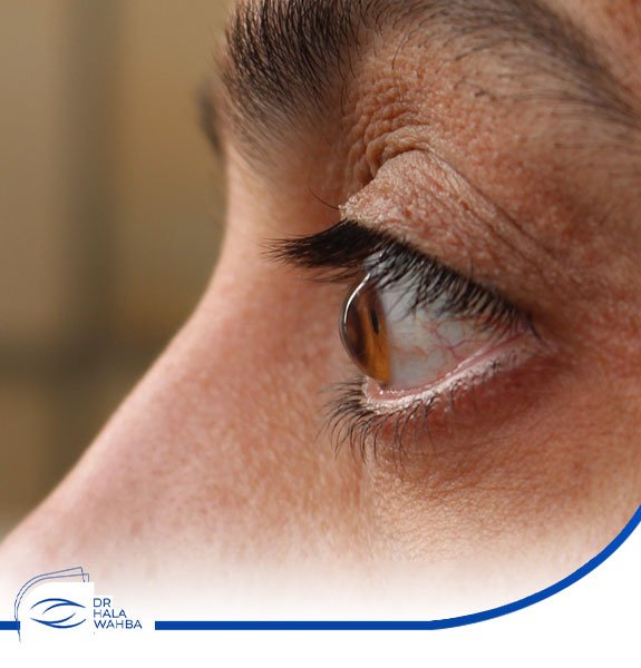
What is keratoconus?
Keratoconus is one of the most common and common eye diseases and is a condition that causes a weakened cornea that is normally round in shape to deviate into an irregular or conical shape.
This irregular shape affects the light and results in its refraction several times while entering the eye on its way to the retina, causing blurring and blurring of vision. Keratoconus occurs in one or both eyes and develops slowly. This disease is common in the age group between 10- 25 years old.
Symptoms of keratoconus
The change in the shape of the cornea to an irregular or conical shape that greatly affects vision and causes blurring of vision. Symptoms associated with keratoconus include:
The patient sees dark shadows.
Extreme sensitivity to light.
Blurred and blurred vision.
In advanced cases of keratoconus, the use of contact lenses becomes uncomfortable and less effective, as the shape of the cornea becomes like a cone and is very clear.
Eye swelling and redness.
Constant change in vision.
Causes of keratoconus
There are no specific causes for this condition, and the factors that contribute to the occurrence of keratoconus include:
Some studies have indicated that keratoconus occurs due to an imbalance in the enzymes that are found inside the cornea, and this increases the possibility of damage to the cornea, as a result of oxidation of some of the compounds present in it, which causes the conical shape of the cornea.
Some scientists believe that keratoconus is due to genetic causes and may be associated with genetic diseases such as Down syndrome. Family history may be one of the main causes of keratoconus.
Excessive exposure to ultraviolet rays in addition to frequent eye rubbing.
Excessive use of contact lenses for long periods may cause what is called keratoconus.
Complications of keratoconus
Sometimes keratoconus may develop and cause some complications, such as swelling of the cornea, which causes a sudden decrease in vision and this requires surgery.
Diagnosis of keratoconus
When visiting the doctor, he examines the patient’s eye by measuring the curvature of the cornea. In some cases, many different tests may be performed to obtain a proper diagnosis of the condition of the eye. The tests for diagnosing this condition include the following:
Corneal topography examination:
This examination is one of the most famous medical examinations used to detect keratoconus. It is an imaging of the cornea and the construction of a topographic map of the surface of the cornea. The map shows with relative accuracy the curvature of the cornea at each point, and the color table shows the different convexities.
In the normal cornea, the convexities are uniform, and therefore the color of the terrain is also uniform. In corneas conical, one area can be seen with very many convexities and this region is marked in orange or red.
This examination is done by placing the patient’s head against the device, and within about a minute, a color image of the cornea is obtained, and through this examination, the curvature of the cornea can be carefully monitored.
Corneal thickness test:
This examination is based on corneal ultrasound, as this examination gives a measurement of the thickness of the cornea at different points, for each person has a different thickness of the cornea, the average thickness of the cornea in humans is about 350 microns, and for people with keratoconus, the thickness of the cornea is less than 500 microns and in advanced cases Of the disease the thickness is less than 400 microns.
Slit lamp examination:
This examination is a microscope-based device that allows examining the eye at very high magnification. In cases of advanced keratoconus, fine lines parallel to each other can be seen on the inner part of the cornea, as well as sometimes scarring in the center of the cornea.
Keratoconus treatment
Keratoconus is treated in several stages. It also depends on several factors, such as its severity, the degree of disease development at the time of diagnosis, and the patient’s ability to see. The following treatment methods are:
There are many treatment methods for keratoconus, including:
the glasses
In the early stages of keratoconus, eyeglasses may replace operations or other procedures in correcting some visual problems, such as myopia, for example.
But glasses do not stop the conic deviation from increasing, so they are a temporary solution.
hard eye lenses
Rigid contact lenses are contact lenses that are similar to regular contact lenses, but differ only in their hardness.
Ordinary lenses are flexible and soft, while lenses that are used in the treatment of keratoconus are rigid.
To strengthen and tighten the surface of the cornea and return it to its natural curvature.
Rigid lenses are also considered a temporary solution, because they only correct vision problems, but they do not stop the aberration or prevent the progression of the curvature.
Crosslinking corneal fixation
Corneal stabilization is the first method of treating the disease, stopping the convexity and increasing the hardness of the cornea.
This process is done by placing vitamin B2 on the cornea and then shedding a strong dose of ultraviolet rays on it.
The triple interaction between corneal tissue, vitamin and ultraviolet rays results in an increase in corneal stiffness and stability and the cessation of cone aberration.
INTACS Ring زراعة
Another method used in the treatment of keratoconus is the process of ring implantation, in which ring stents are installed to adjust the degree of curvature or curvature of the cornea and stabilize it.
Cornea Transplant
In late cases that cannot be treated with rigid lenses or through fixations;
Corneal transplantation is resorted to, and corneal transplantation in Egypt is one of the most successful operations recently
Due to the decreasing possibility of the body rejecting the transplanted cornea after the tremendous scientific developments that occurred in the field of ophthalmology,
which greatly increased the odds of success,